missing translation for 'onlineSavingsMsg'
Learn More
Learn More
Perforin Monoclonal Antibody (eBioOMAK-D), eBioscience™, Invitrogen™
Rat Monoclonal Antibody
Brand: Invitrogen 14-9392-80
This item is not returnable.
View return policy
Description
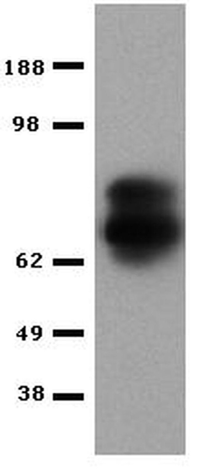
Description: The eBioOMAK-D antibody reacts with mouse perforin (pore-forming protein, pfp, Prf). Perforin is one of the cytolytic mediators present in the cytoplasmic granules of cytotoxic T lymphocytes (CTL) and natural killer cells (NK). Perforin is involved in the killing function by CTLs and NKs and has an important role in the immune response against tumors and virus infections. By immunobloting, eBioOMAK-D recognizes a ∽70kDa band in lysates of CTLL-2 mouse cytotoxic cell line and in lysates of IL-2 stimulated but not unstimulated mouse splenocytes. By multi-color intracellular flow cytometric analysis, eBioOMAK-D staining is increased upon stimulation (IL-2 or anti-CD3/28). Intracellular flow staining results showing upregulation of protein expression have been confirmed by immunoblotting. Furthermore, stimulated Perforin Knock-out (developed by Walsh) splenocytes do not stain with eBioOMAK-D nor is any protein detectable by western blotting with eBioOMAK-D as well as other anti-mouse perforin antibodies. Please note that the Kagi perforin knock-out mice may synthesize a truncated form of the protein which may be recognized by eBioOMAK-D. In IL-2 stimulated mouse splenocytes, NK cells (as determined by CD49b staining) contain perforin while CD8 cells contain little to none and can vary with culture conditions. This has been confirmed by staining and western blotting the two populations using both OMAK-D and P1-8 antibodies.
In contrast stimulation of splenocytes with anti-CD3/CD28 antibodies does result in an increase of perforin on both NK cells and CD8 cells. eBioOMAK-D is also crossreactive to human perforin and co-stains CD56 positive cells in PBMC. Expression of perforin and Granzyme B do not always correlate (as discussed above in the CD8 population of IL-2 stimulated splenocytes). Granzyme B typically is expressed earlier and at higher levels. Expression of Granzyme B is dramatically increased (more than 10,00 fold based on mRNA estimates and significantly at the protein level based on western blotting and flow analysis) compared to a minimal increase (10-100 fold) in perforin mRNA and protein with IL-2 stimulation. For intracellular staining and flow cytometric analysis with direct conjugates of anti-mouse perforin, it is highly recommended to use the Foxp3 buffer system (cat. 00-5523). Other buffers may yield varying results. For more information, please contact technical support at tech@ebioscience.com. Applications Reported: This eBioOMAK-D antibody has been reported for use in immunoblotting (WB). Applications Tested: This eBioOMAK-D antibody has been tested by immunblotting of CTLL-2 cell lysates and can be used at less than or equal to 2 μg/mL. Purity: Greater than 90%, as determined by SDS-PAGE. Aggregation: Less than 10%, as determined by HPLC. Filtration: 0.2 μm post-manufacturing filtered. Perforin is one of the major cytolytic proteins of cytolytic granules. Perforin is a cytolytic mediator and is stored in and released by cytoplasmic granules. Moreover, perforin is involved in immune defense against tumors and virus infections as mediated by cytotoxic lymphocytes. Perforin is a 555 amino acid protein with a 21 amino acid signal peptide, and has a molecular weight of 70 to 75 kD. Perforin is a pore forming protein with a mechanism of transmembrane channel formation similar to C9, and homology between perforin and C9 have been demonstrated. Studies show that perforin is expressed only in killer cell lines and not in helper T lymphocytes or other tumor cells tested. Perforin is known to be a key effector molecule for T-cell- and natural killer-cell-mediated cytolysis. Defects in the perforin gene cause familial hemophagocytic lymphohistiocytosis type 2 (HPLH2), a rare and lethal autosomal recessive disorder of early childhood. Alternative splicing results in multiple transcript variants of perforin.Specifications
| Perforin | |
| Monoclonal | |
| 0.5 mg/mL | |
| PBS with 0.09% sodium azide; pH 7.2 | |
| P10820 | |
| Prf1 | |
| Affinity chromatography | |
| RUO | |
| 18646 | |
| 4° C | |
| Liquid |
| Western Blot | |
| eBioOMAK-D | |
| Unconjugated | |
| Prf1 | |
| cytolysin; FLH2; HPLH2; lymphocyte pore forming protein; lymphocyte pore-forming protein; perforin 1 (pore forming protein); perforin-1; PFN1; PFP; RP11-710A11.3 | |
| Rat | |
| 25 μg | |
| Primary | |
| Mouse | |
| Antibody | |
| IgG2a κ |
Product Content Correction
Your input is important to us. Please complete this form to provide feedback related to the content on this product.
Product Title
For Research Use Only.
Spot an opportunity for improvement?Share a Content Correction