missing translation for 'onlineSavingsMsg'
Learn More
Learn More
Invitrogen™ CD93 (AA4.1) Monoclonal Antibody (R139), eBioscience™

Mouse Monoclonal Antibody
£192.00
Specifications
| Antigen | CD93 (AA4.1) |
|---|---|
| Clone | R139 |
| Concentration | 0.5 mg/mL |
| Content And Storage | 4°C |
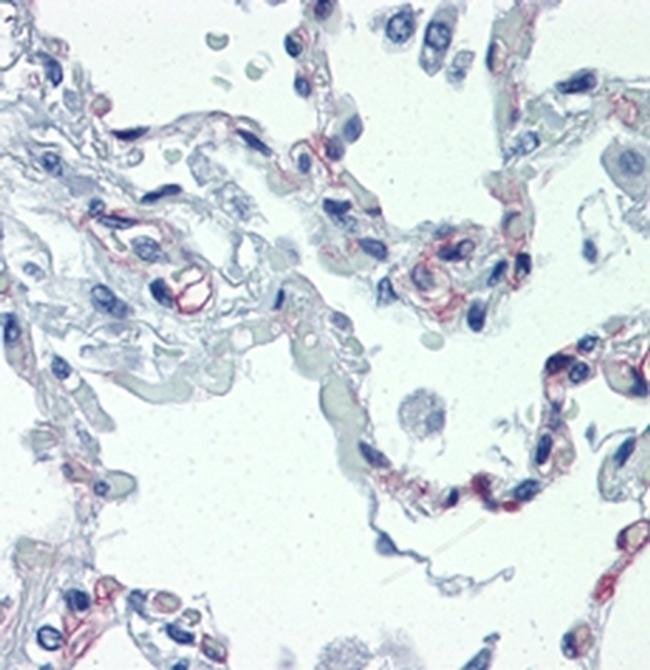
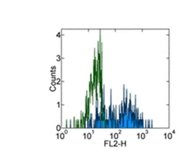
| Applications | ELISA, Flow Cytometry, Immunohistochemistry (Paraffin), Immunoprecipitation, Western Blot |
Description
Description: The monoclonal antibody R139 recognizes human CD93, also known as C1qRp. The glycoprotein CD93 binds to C1q, the subunit of the complement protein, mannose binding lectin and pulmonary surfactant protein A. CD93 is predicted to play a role in the clearance of apoptotic cells. Expression of CD93 is confined to myeloid cells with higher expression on monocytes than neutrophils, eosinophils, platelets and endothelial cells. Expression on DC's is downregulated upon maturation. Additionally, CD93 has been shown to define an early bone marrow stem cell population of hematopoietic and hepatic precursors. The monoclonal antibody R139 blocks C1q-mediated enhancement of phagocytosis. CD93 can be shed from the cell surface. This phenomenon can be measured using R3 antibody as detection with R139 as capture to detect soluble CD93 by ELISA. The epitope for R139 resides in the EGF domains. Applications Reported: This R139 antibody has been reported for use in flow cytometric analysis, immunoprecipitation, immunoblotting (WB) under nonreducing conditions, ELISA, and immunohistochemistry of formalin-fixed paraffin embedded tissue. Applications Tested: This R139 antibody has been tested by flow cytometric analysis of human peripheral blood cells. This can be used at less than or equal to 0.5 μg per test. A test is defined as the amount (μg) of antibody that will stain a cell sample in a final volume of 100 μL. Cell number should be determined empirically but can range...
The protein encoded by this gene is a cell-surface glycoprotein and type I membrane protein that was originally identified as a myeloid cell-specific marker. The encoded protein was once thought to be a receptor for C1q, but now is thought to instead be involved in intercellular adhesion and in the clearance of apoptotic cells. The intracellular cytoplasmic tail of this protein has been found to interact with moesin, a protein known to play a role in linking transmembrane proteins to the cytoskeleton and in the remodelling of the cytoskeleton.Specifications
| CD93 (AA4.1) | |
| 0.5 mg/mL | |
| ELISA, Flow Cytometry, Immunohistochemistry (Paraffin), Immunoprecipitation, Western Blot | |
| Unconjugated | |
| Mouse | |
| RUO | |
| PBS with 0.09% sodium azide; pH 7.2 | |
| Q9NPY3 | |
| 22918 | |
| Primary | |
| Affinity chromatography |
| R139 | |
| 4°C | |
| Monoclonal | |
| Liquid | |
| IgG2b | |
| Human | |
| Cd93 | |
| 6030404G09Rik; AA145088; Aa4; AA4.1; AW555904; C1q receptor; C1q receptor 1; C1q/MBL/SPA receptor; C1qR; C1qR(P); C1qr1; C1qRP; Cd93; CD93 antigen; CD93 molecule; CDw93; Cell surface antigen AA4; complement component 1 q subcomponent receptor 1; complement component 1, q subcomponent, receptor 1; complement component C1q receptor; dJ737E23.1; ECSM3; Ly68; ly-68; Lymphocyte antigen 68; matrix-remodeling-associated protein 4; matrix-remodelling associated 4; MXRA4 | |
| Cd93 | |
| Antibody |
Spot an opportunity for improvement?Share a Content Correction
Product Content Correction
Your input is important to us. Please complete this form to provide feedback related to the content on this product.
Product Title