missing translation for 'onlineSavingsMsg'
Learn More
Learn More
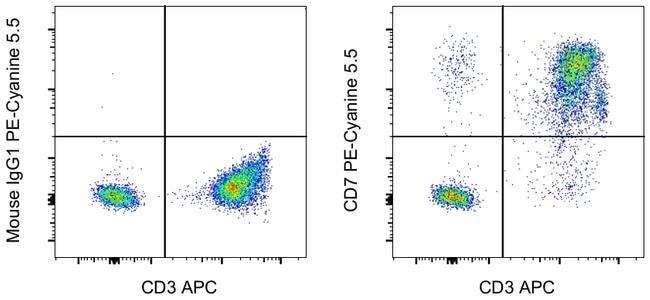
Invitrogen™ CD7 Monoclonal Antibody (eBio124-1D1 (124-1D1)), PE-Cyanine5.5, eBioscience™

Mouse Monoclonal Antibody
£315.00
Specifications
| Antigen | CD7 |
|---|---|
| Clone | eBio124-1D1 (124-1D1) |
| Concentration | 5 μL/Test |
| Content And Storage | 4°C, store in dark, DO NOT FREEZE! |
| Applications | Flow Cytometry |
Description
Description: The eBio124-1D1 monoclonal antibody reacts with human CD7, also known as gp40 and Leu9. CD7, a 40 kD receptor, is a member of the immunoglobulin gene superfamily. The N-terminal amino acid sequence (aa1-107) is highly homologous to Ig kappa light chain sequence; while the carboxyl-terminal region of the extracellular domain is proline-rich and has been postulated to form a stalk from which the Ig domain projects. CD7 is expressed on the majority of immature and mature T lymphocytes, and T cell leukemias. It is also found on natural killer cells, a small suppopulation of normal B cells and on maligant B cells. Cross-linking surface CD7 positively modulates T cell and NK cell activity, as measured by calcium flux, expression of adhesion molecules, cytokine secretion and proliferation. CD7 associates directly with phosphoinositol 3'-kinase. CD7 ligation induces production of D-3 phosphoinositides and tyrosine phosphorylation. A clonogenic subpopulation of human CD34(+) CD38(-) cord blood cells that express CD45RA and HLA-DR and high levels of the CD7 has been reported. These cells possess the capacity for lymphopoiesis. They can generate B-cells, natural killer cells, and dendritic cells but do not possess the capacity to develop into myeloid cells or erythroid cells. The CD7(+) phenotype distinguishes primitive human lymphoid progenitors from pluripotent stem cells. Furthermore, it has been suggested that CD7 co-operates with CD28 during Treg function, as mice d...
CD7, also known as gp40 or Leu9, is a 40 kDa receptor and member of the immunoglobulin gene superfamily. It features an N-terminal region (amino acids 1-107) that is highly homologous to Ig kappa light chains, while its carboxyl-terminal region is proline-rich, forming a stalk from which the Ig domain projects. CD7 is prominently expressed on the majority of immature and mature T lymphocytes, as well as T cell leukemias. It is also found on natural killer cells, a small subpopulation of normal B cells, and malignant B cells. CD7 plays a crucial role in modulating immune cell activity. Cross-linking of surface CD7 enhances T cell and NK cell functions, as evidenced by increased calcium flux, expression of adhesion molecules, cytokine secretion, and proliferation. CD7 directly associates with phosphoinositol 3-kinase, and its ligation induces the production of D-3 phosphoinositides and tyrosine phosphorylation. The expression of CD7 is an important marker in leukemia diagnostics, highlighting its significance in both normal immune function and disease states.Specifications
| CD7 | |
| 5 μL/Test | |
| Flow Cytometry | |
| PE-Cyanine5.5 | |
| Mouse | |
| RUO | |
| PBS with BSA and 0.09% sodium azide; pH 7.2 | |
| P09564 | |
| 924 | |
| Primary | |
| Affinity chromatography |
| eBio124-1D1 (124-1D1) | |
| 4°C, store in dark, DO NOT FREEZE! | |
| Monoclonal | |
| Liquid | |
| IgG1 κ | |
| Human | |
| CD7 | |
| Cd7; CD7 antigen; CD7 antigen (p41); Cd7 molecule; GP40; LEU-9; p41 protein; T-cell antigen CD7; T-cell leukemia antigen; T-cell surface antigen Leu-9; Tp40; TP41 | |
| CD7 | |
| Antibody |
Spot an opportunity for improvement?Share a Content Correction
Product Content Correction
Your input is important to us. Please complete this form to provide feedback related to the content on this product.
Product Title