missing translation for 'onlineSavingsMsg'
Learn More
Learn More
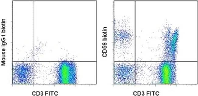
Invitrogen™ CD56 (NCAM) Monoclonal Antibody (CMSSB), Biotin, eBioscience™

Mouse Monoclonal Antibody
£171.00
Specifications
| Antigen | CD56 (NCAM) |
|---|---|
| Clone | CMSSB |
| Concentration | 0.5 mg/mL |
| Content And Storage | 4°C, store in dark, DO NOT FREEZE! |
| Applications | Flow Cytometry, Immunohistochemistry (Paraffin) |
Description
Description: This CMSSB monoclonal antibody reacts with human CD56, also known as Neural Cell Adhesion Molecule (NCAM). CD56 is a highly glycosylated transmembrane molecule expressed by neurons and plays a role in the homotypic adhesion of neural cells. In the hematopoietic system, CD56 is expressed on NK cells and a subset of T cells referred to as NKT cells. Staining with CMSSB does not block binding of MEM188 or CB56. Applications Reported: This CMSSB antibody has been reported for use in flow cytometric analysis and immunohistology staining of paraffin embedded tissue sections. Applications Tested: This CMSSB antibody has been tested by flow cytometric analysis of normal human peripheral blood cells. This can be used at less than or equal to 0.5 μg per test. A test is defined as the amount (μg) of antibody that will stain a cell sample in a final volume of 100 μL. Cell number should be determined empirically but can range from 10^5 to 10^8 cells/test. It is recommended that the antibody be carefully titrated for optimal performance in the assay of interest. Filtration: 0.2 μm post-manufacturing filtered.
CD56, also known as neural cell adhesion molecule (NCAM), is a highly glycosylated transmembrane glycoprotein of the immunoglobulin family. It plays a crucial role in cell adhesion, migration, axonal growth, pathfinding, and synaptic plasticity. CD56 is ubiquitously expressed in the nervous system in isoforms ranging from 120-180 kDa and is involved in homotypic adhesion of neural cells. It mediates interactions by binding extracellular matrix components such as laminin and integrins, with polysialic modification reducing CD56-mediated adhesion. In the hematopoietic system, CD56 is expressed on natural killer (NK) cells and a subset of T cells known as NKT cells. It is also found on most neuroectodermal-derived cell lines, tissues, and neoplasms, including retinoblastoma, medulloblastoma, astrocytomas, and neuroblastoma. CD56 serves as a widely used neuroendocrine marker with high sensitivity for neuroendocrine tumors and ovarian granulosa cell tumors. Diseases associated with CD56 dysfunction include rabies and blastic plasmacytoid dendritic cell neoplasms, highlighting its importance in both neural and immune system functions.Specifications
| CD56 (NCAM) | |
| 0.5 mg/mL | |
| Flow Cytometry, Immunohistochemistry (Paraffin) | |
| Biotin | |
| Mouse | |
| RUO | |
| PBS with 0.09% sodium azide; pH 7.2 | |
| P13591 | |
| 4684 | |
| Primary | |
| Affinity chromatography |
| CMSSB | |
| 4°C, store in dark, DO NOT FREEZE! | |
| Monoclonal | |
| Liquid | |
| IgG1 κ | |
| Human | |
| Ncam1 | |
| adhesion molecule; antigen recognized by monoclonal antibody 5.1H11; Cd56; CD-56; CD56 120 kDa GPI-linked isoform; CD56 140 kDa isoform; CD56 140 kDa VASE isoform; E NCAM; E-NCAM; MSK39; N CAM1; NCAM; N-CAM; Ncam1; N-CAM-1; NCAM-1; NCAMC; NCAM-C; neural cell adhesion molecule; Neural cell adhesion molecule 1; neural cell adhesion molecule, NCAM; sCD56; sNCAM; soluble CD56; soluble NCAM | |
| Ncam1 | |
| Antibody |
Spot an opportunity for improvement?Share a Content Correction
Product Content Correction
Your input is important to us. Please complete this form to provide feedback related to the content on this product.
Product Title