missing translation for 'onlineSavingsMsg'
Learn More
Learn More
CD43/Sialophorin Antibody (DF-T1), Novus Biologicals™
Mouse Monoclonal Antibody
£232.00 - £462.00
Specifications
| Antigen | CD43/Sialophorin |
|---|---|
| Clone | DF-T1 |
| Concentration | 0.2mg/mL |
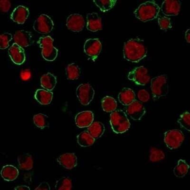
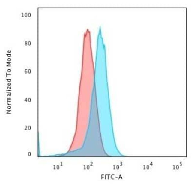
| Dilution | Western Blot 0.5-1ug/ml, Flow Cytometry 0.5-1ug/million cells, ELISA 1-5ug/ml for coating, Immunocytochemistry/Immunofluorescence 1-2ug/ml, Immunoprecipitation 1-2ug/500ug protein lysate, Immunohistochemistry-Paraffin 0.5-1ug/ml, Immunohistochemistry-Frozen 0.5-1ug/ml |
| Classification | Monoclonal |
| Product Code | Brand | Quantity | Price | Quantity & Availability | |||||
|---|---|---|---|---|---|---|---|---|---|
| Product Code | Brand | Quantity | Price | Quantity & Availability | |||||
|
18198223
|
Novus Biologicals
NBP2-15190-0.1MG |
0.1 mg |
£392.00
0.10mg |
Please sign in to purchase this item. Need a web account? Register with us today! | |||||
|
18191644
|
Novus Biologicals
NBP2-15190-0.2MG |
0.2 mg |
£462.00
0.20mg |
Please sign in to purchase this item. Need a web account? Register with us today! | |||||
|
18449871
|
Novus Biologicals
NBP2-15190-20ug |
20 ug |
£232.00
20µg |
Please sign in to purchase this item. Need a web account? Register with us today! | |||||
Description
CD43/Sialophorin Monoclonal specifically detects CD43/Sialophorin in Human samples. It is validated for Western Blot, Flow Cytometry, Immunohistochemistry, Immunocytochemistry/Immunofluorescence, Immunohistochemistry-Paraffin.Specifications
| CD43/Sialophorin | |
| 0.2mg/mL | |
| Monoclonal | |
| Purified | |
| RUO | |
| PBS with 0.05% BSA. with 0.05% Sodium Azide | |
| CD43 antigen, CD43), Galactoglycoprotein, GALGP, Leukocyte sialoglycoprotein, Sialophorin, sialophorin (gpL115, leukosialin, CD43) | |
| SPN | |
| IgG1 κ | |
| Protein G purified | |
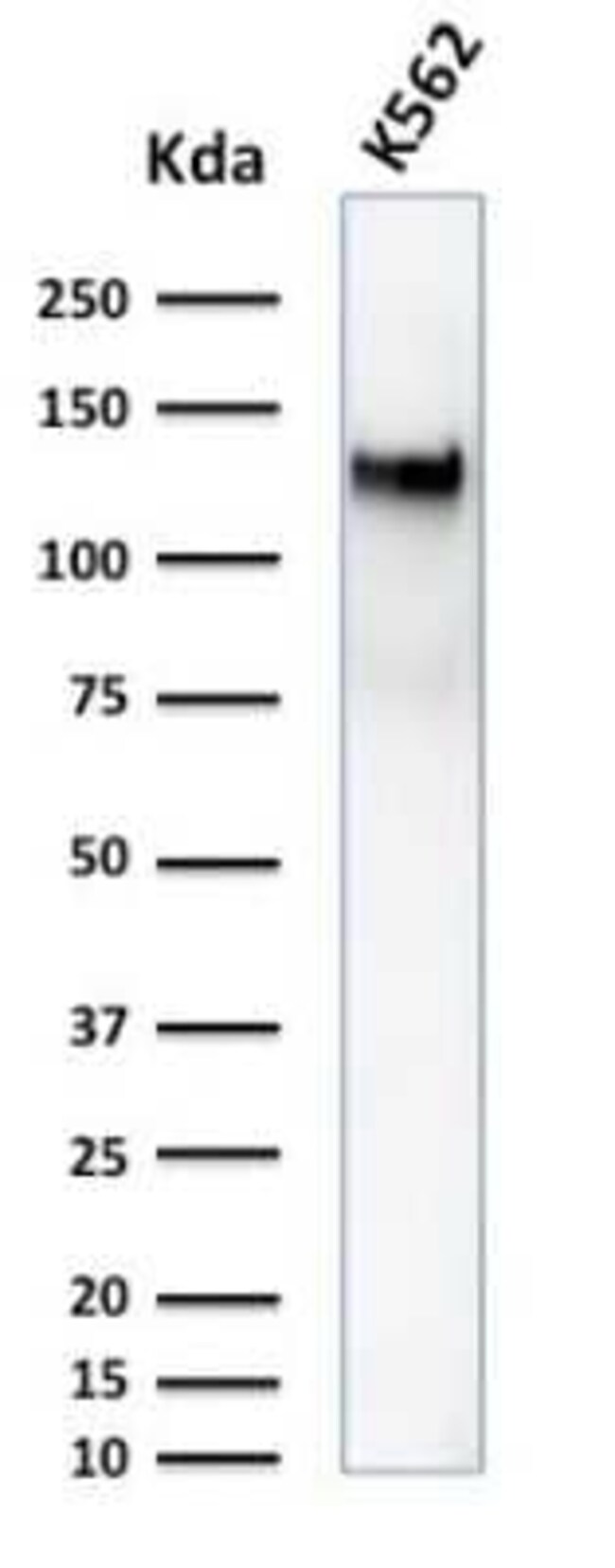
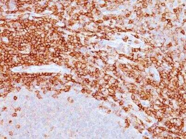
| It recognizes a cell surface glycoprotein of 95/115/135kDa (depending upon the extent of glycosylation), identified as CD43. 70-90% of T-cell lymphomas and from 22-37% of B-cell lymphomas express CD43. No reactivity has been observed with reactive B-cells. So a B-lineage population that co-expresses CD43 is highly likely to be a malignant lymphoma, especially a low-grade lymphoma, rather than a reactive B-cell population. When CD43 antibody is used in combination with anti-CD20, effective immunophenotyping of the lymphomas in formalin-fixed tissues can be obtained. Co-staining of a lymphoid infiltrate with anti-CD20 and anti-CD43 argues against a reactive process and favors a diagnosis of lymphoma. |
| DF-T1 | |
| Western Blot 0.5-1ug/ml, Flow Cytometry 0.5-1ug/million cells, ELISA 1-5ug/ml for coating, Immunocytochemistry/Immunofluorescence 1-2ug/ml, Immunoprecipitation 1-2ug/500ug protein lysate, Immunohistochemistry-Paraffin 0.5-1ug/ml, Immunohistochemistry-Frozen 0.5-1ug/ml | |
| Unconjugated | |
| Mouse | |
| B Cell Development and Differentiation Markers, Immunology | |
| P16150 | |
| 6693 | |
| Myeloblastic KG1 cells were used as the immunogen for this antibody. | |
| Primary | |
| Store at 4C. |
For Research Use Only
Spot an opportunity for improvement?Share a Content Correction
Product Content Correction
Your input is important to us. Please complete this form to provide feedback related to the content on this product.
Product Title