missing translation for 'onlineSavingsMsg'
Learn More
Learn More
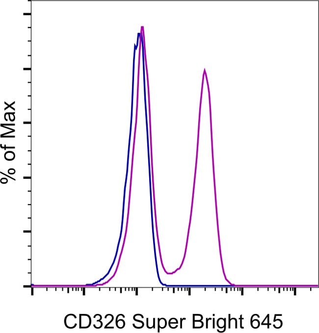

CD326 (EpCAM) Monoclonal Antibody (G8.8), Super Bright™ 645, eBioscience™, Invitrogen™

Rat Monoclonal Antibody
£130.00 - £337.00
Specifications
| Antigen | CD326 (EpCAM) |
|---|---|
| Clone | G8.8 |
| Concentration | 0.2 mg/mL |
| Applications | Flow Cytometry |
| Classification | Monoclonal |
Description
Description: The eBioC1.7 monoclonal antibody reacts with human CD244 (2B4, p38). In human, CD244 is a 38 kDa protein expressed on NK cells, a subset of CD8+ T cells, gammadelta T cells, monocytes, basophils and eosinophils. Binding of the CD244 ligand, CD48, results in NK cell activation, unlike mouse CD244, which is an inhibitory receptor. For CD244 expressed on NK cells, binding of CD48 results in enhanced NK cell cytotoxicity and secretion of IFN-gamma. Recently, it was demonstrated that binding of the C1.7 monoclonal antibody and CD48 involve the same residue in the V domain of human CD244, which explains the ability of C1.7 binding to induce activation of NK cells. Binding of C1.7 to CD244 leads to tyrosine phosphorylation and recruitment of the adaptor molecule SAP (SLAM-associated protein). Patients with X-linked lymphoproliferative disorder (XLPD) have a mutation in SAP which renders it unable to bind to phosphorylated CD244. Applications Reported: This eBioC1.7 antibody has been reported for use in flow cytometric analysis. Applications Tested: This eBioC1.7 antibody has been pre-diluted and tested by flow cytometric analysis of normal human peripheral blood cells. This may be used at 5 μL (1 μg/mL) per test. A test is defined as the amount (μg) of antibody that will stain a cell sample in a final volume of 100 μL. Cell number should be determined empirically but can range from 10^5 to 10^8 cells/test.
Ep-CAM (epithelial adhesion molecule, epithelial specific antigen, ESA) is a transmembrane glycoprotein expressed in the epithelium with a molecular weight of approximately 40 kDa, which functions as an epithelial cell adhesion molecule. Ep-CAM functions as a homotypic calcium-independent cell adhesion molecule, and has a direct impact on cell cycle, proliferation and metabolism of epithelial cells and fibroblasts due to its ability to rapidly induce the proto-oncogene c-myc and the cell cycle regulating genes cyclin A and E. Ep-CAM mediates Ca2+-independent homotypic interactions. Formation of Ep-CAM-mediated adhesions have a negative regulatory effect on adhesions mediated by classic cadherins, which may have strong effects on the differentiation and growth of epithelial cells. Ep-CAM overexpression was suggested to be associated with enhanced epithelial proliferation. Ep-CAM is highly expressed in human carcinomas, and is a marker for tumors of epithelial lineage. Ep-CAM is expressed on baso-lateral cell surface in most simple epithelia and many carcinoma types. Also, Ep-CAM reportedly distinguishes adenocarcinomas from pleural mesotheliomas.Specifications
| CD326 (EpCAM) | |
| 0.2 mg/mL | |
| Monoclonal | |
| Liquid | |
| RUO | |
| Q99JW5 | |
| IgG2a κ | |
| Affinity chromatography | |
| Antibody |
| G8.8 | |
| Flow Cytometry | |
| Super Bright 645 | |
| Rat | |
| Mouse | |
| 17075 | |
| Primary | |
| 4° C, store in dark, DO NOT FREEZE! |
Spot an opportunity for improvement?Share a Content Correction
Product Content Correction
Your input is important to us. Please complete this form to provide feedback related to the content on this product.
Product Title