missing translation for 'onlineSavingsMsg'
Learn More
Learn More
CD223 (LAG-3) Monoclonal Antibody (eBioC9B7W (C9B7W)), Super Bright™ 600, eBioscience™, Invitrogen™
Rat Monoclonal Antibody
Brand: eBioscience 63-2231-80
This item is not returnable.
View return policy
Description
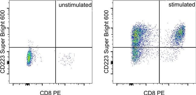
Description: The eBioC9B7W monoclonal antibody recognizes mouse CD223 (LAG-3, LAG3) protein expressed by activated alpha/beta-TCR bearing T cells, a subset of gamma/delta-TCR bearing T cells and a subset of NK cells. CD223 is a 70 kDa type I transmembrane protein with a structure that is similar to CD4. However, a soluble form of human CD223 has been detected by ELISA in human serum, and data suggest that mouse CD223 is proteolytically cleaved in the D4 domain. This results in a 54 kDa fragment containing all the extracellular domains, and a 16 kDa fragment containing the intracellular and transmembrane domains. The 54 kDa can remain membrane-associated or be released as soluble CD223. CD223 binds to MHC class II with higher affinity than CD4, and it is thought that this interaction is involved in the negative regulation of T-cell activation and homeostatic proliferation. Furthermore, CD223 is expressed by CD4+CD25+ regulatory T cells, and it has been suggested that CD223 may be involved in their regulatory function. Applications Reported: This eBioC9B7W antibody has been reported for use in flow cytometric analysis. Applications Tested: This eBioC9B7W antibody has been tested by flow cytometric analysis of stimulated mouse splenocytes. This may be used at less than or equal to 1.0 μg per test. A test is defined as the amount (μg) of antibody that will stain a cell sample in a final volume of 100 μL.
Cell number should be determined empirically but can range from 10^5 to 10^8 cells/test. It is recommended that the antibody be carefully titrated for optimal performance in the assay of interest. Super Bright 600 is a tandem dye that can be excited with the violet laser line (405 nm) and emits at 600 nm. We recommend using a 610/20 bandpass filter. Please make sure that your instrument is capable of detecting this fluorochrome. When using two or more Super Bright dye-conjugated antibodies in a staining panel, it is recommended to use Super Bright Complete Staining Buffer (Product No. SB-4401) to minimize any non-specific polymer interactions. Please refer to the datasheet for Super Bright Staining Buffer for more information. Light sensitivity: This tandem dye is sensitive to photo-induced oxidation. Please protect this vial and stained samples from light. Fixation: Samples can be stored in IC Fixation Buffer (Product No. 00-8222) (100 μL of cell sample + 100 μL of IC Fixation Buffer) or 1-step Fix/Lyse Solution (Product No. 00-5333) for up to 3 days in the dark at 4°C with minimal impact on brightness and FRET efficiency/compensation. Some generalizations regarding fluorophore performance after fixation can be made, but clone specific performance should be determined empirically. Excitation: 405 nm; Emission: 600 nm; Laser: Violet Laser Super Bright Polymer Dyes are sold under license from Becton, Dickinson and Company. LAG-3 is a 70-kDa surface glycoprotein belonging to the Ig superfamily with homology to CD4. LAG-3 binds to MHC class II with higher affinity than CD4 and is thought to be involved in the negative regulation of T cell activation and homeostatic proliferation. Surface expression of LAG-3 has been reported on activated T cells (including regulatory T cells) and NK cells. CD8+ T cells usually express LAG-3 at significantly higher levels than CD4+ T cells. Coexpression of LAG-3 and CD49b has been proposed to identify human and mouse Type 1 regulatory T cells (Tr1 cells).Specifications
| CD223 (LAG-3) | |
| Monoclonal | |
| 0.2 mg/ml | |
| PBS with BSA and 0.09% sodium azide; pH 7.2 | |
| Q61790 | |
| Lag3 | |
| Affinity chromatography | |
| RUO | |
| 16768 | |
| 4° C, store in dark, DO NOT FREEZE! | |
| Liquid |
| Flow Cytometry | |
| eBioC9B7W (C9B7W) | |
| Super Bright 600 | |
| Lag3 | |
| CD223; Ly66; lymphocyte-activation gene 3 | |
| Rat | |
| 25 μg | |
| Primary | |
| Mouse | |
| Antibody | |
| IgG1 κ |
Product Content Correction
Your input is important to us. Please complete this form to provide feedback related to the content on this product.
Product Title
Spot an opportunity for improvement?Share a Content Correction